Results
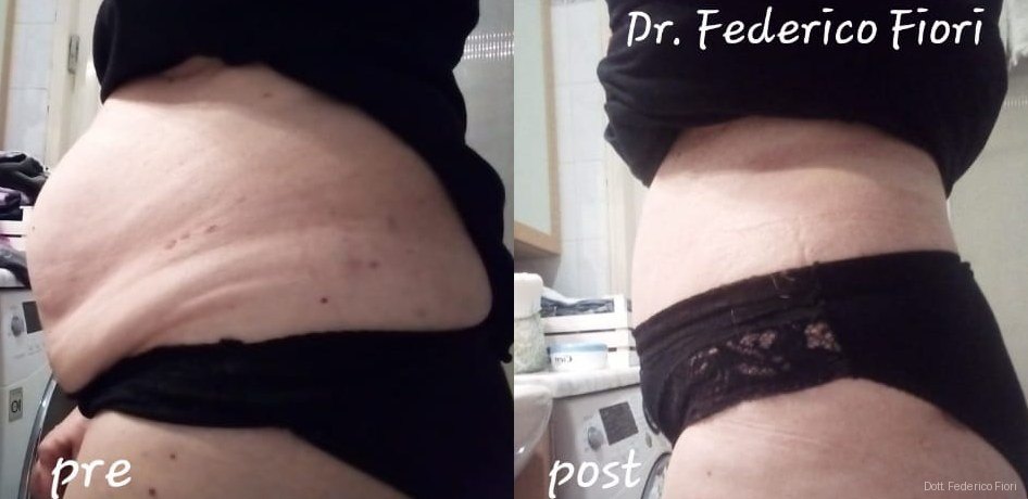
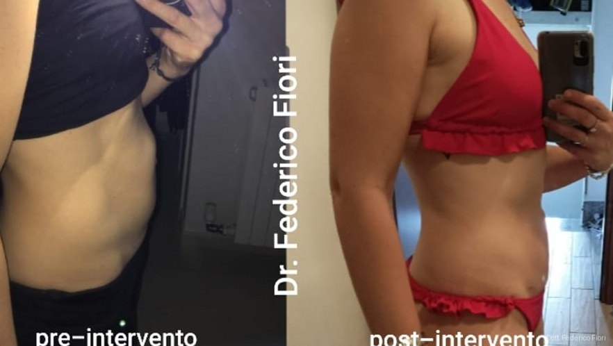
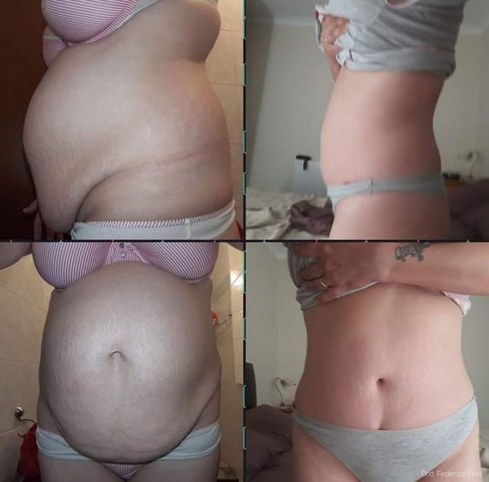
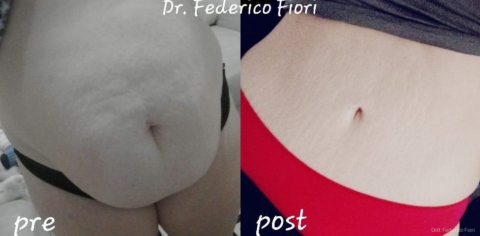
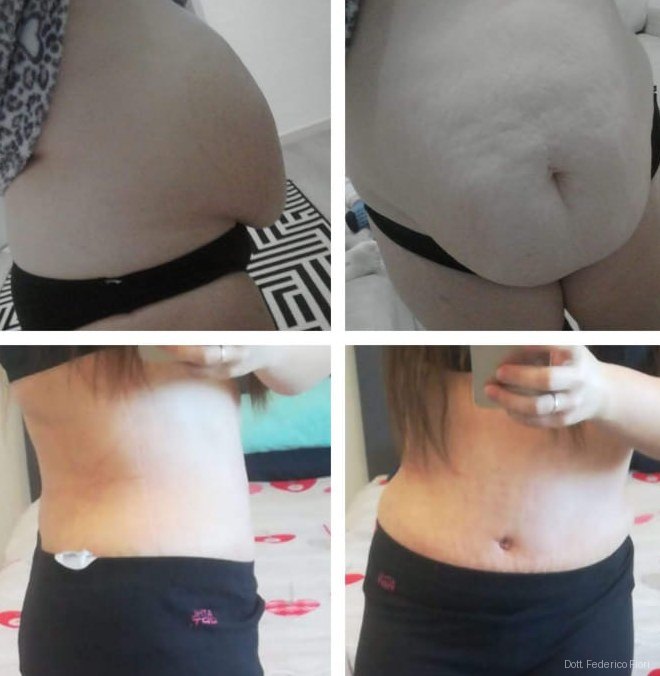
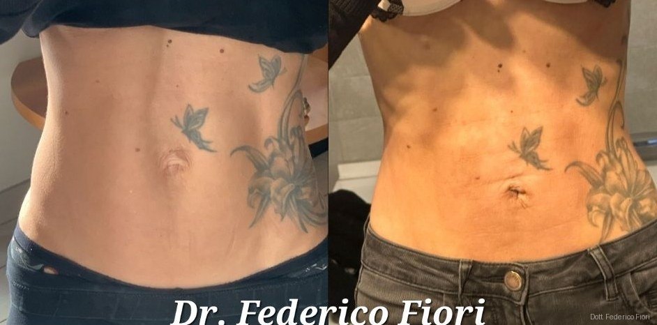
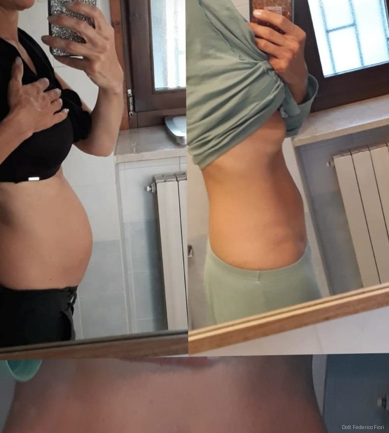
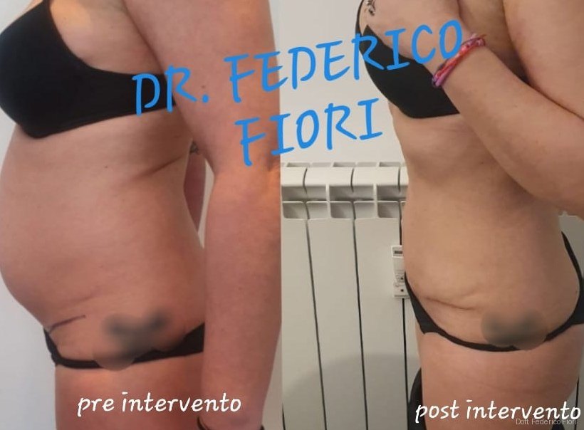
The images document; they do not promise: the same technique on two different people produces two different outcomes.
This page collects pre- and postoperative images of cases I have operated on: rectus diastasis, abdominal wall hernias, incisional hernias, abdominoplasties and complex reconstructions. Each case states the technique used and how many months after surgery the photograph was taken. The images are published with the specific written consent of the people concerned, show no recognizable faces, and do not constitute a prediction of the outcome in other cases.
Why I publish pre and post-operative images
Because anyone deciding whether to have surgery wants to see, not just read. A photograph shows in an instant what changes and what does not, and it lets you come to your consultation with sharper questions. I publish these images under a rule that never changes: always with the person's written consent, always without recognizable faces, always stating the technique and the follow-up interval.
What a photograph can and cannot say
It can show the profile of the abdomen, the reduction in bulging, the position and appearance of the scars, and symmetry. It cannot show the strength of the repair, the restoration of the midline, core function, or pain. For those you need dynamic ultrasound, CT when indicated, and functional testing: these are the tools with which I actually verify the result, and they are the same ones I use to decide whether and how to operate.
Why two similar abdomens give different results
- The starting point: size of the defect, number of previous operations, skin quality;
- Weight and how stable it stays over time: significant weight gain after surgery changes the contour;
- Smoking, blood glucose and nutritional status, which affect wound healing;
- How closely the recovery program is followed, in particular abdominal wall rehabilitation;
- Time: the definitive result is judged at six to twelve months, not at three weeks.
Consent and privacy
Every published image is covered by specific written consent, separate from the consent to surgery and revocable at any time: if someone changes their mind, the image is removed. The photographs are stripped of file metadata, contain no recognizable faces, and identifying features such as tattoos or distinctive moles are masked.
How to read these images
These photographs are clinical documentation, not advertising for a result. The timing of the follow-up changes everything: in the first months postoperative swelling is still present and scars are more visible than they will eventually be. The shape of the abdomen also depends on the starting point, on weight, on tissue quality and on how closely the recovery program is followed. No image can tell you what outcome is achievable in your case: that is established at the consultation, through clinical examination and imaging.
Choose a topic
- Rectus diastasis — In rectus diastasis the post-operative image shows two distinct things: the outer profile of the abdomen and the reconstruction of the midline. The first is visible in a photograph, the second is measured with ultrasound. The images on this page document real cases operated on with endoscopic or open techniques, photographed months after surgery and published with the specific consent of the person. (29)
- Abdominal wall hernias — In abdominal wall hernias — umbilical, epigastric, inguinal — the photograph documents the disappearance of the bulge and respect for the shape of the navel and the natural creases, the detail patients notice first. What a photograph cannot show is how the repair holds over time: that depends on closing the defect, on the plane where the mesh sits and on individual factors. (0)
- Incisional hernia — In incisional hernia the goal is not aesthetic: it is to rebuild a wall that has lost continuity, bringing the rectus muscles back to the midline and giving the abdomen its capacity to contain and to support the trunk again. The images show the reduction of the bulge; the quality of the reconstruction is assessed with CT and with function, not with a photograph. (0)
- Morphological surgery (breast, abdomen, limbs) — Morphological surgery works on shape: abdomen, breast, arms and thighs. Here the photograph is the most direct document, because the result is largely morphological — flank profile, skin tension, symmetry, position of the scars. It should be read knowing that a scar matures over twelve to eighteen months and that in recent images it looks more evident than it will be. (0)
- Complex cases and reconstructions — These are cases with very wide defects, multiple recurrences or the consequences of previous operations, where the wall must be rebuilt in several planes and sometimes with the contribution of plastic surgery. Here the images serve mainly to show that a compromised situation can be taken up again, not to promise the same outcome to anyone. (0)
Rectus diastasis
Rectus diastasis
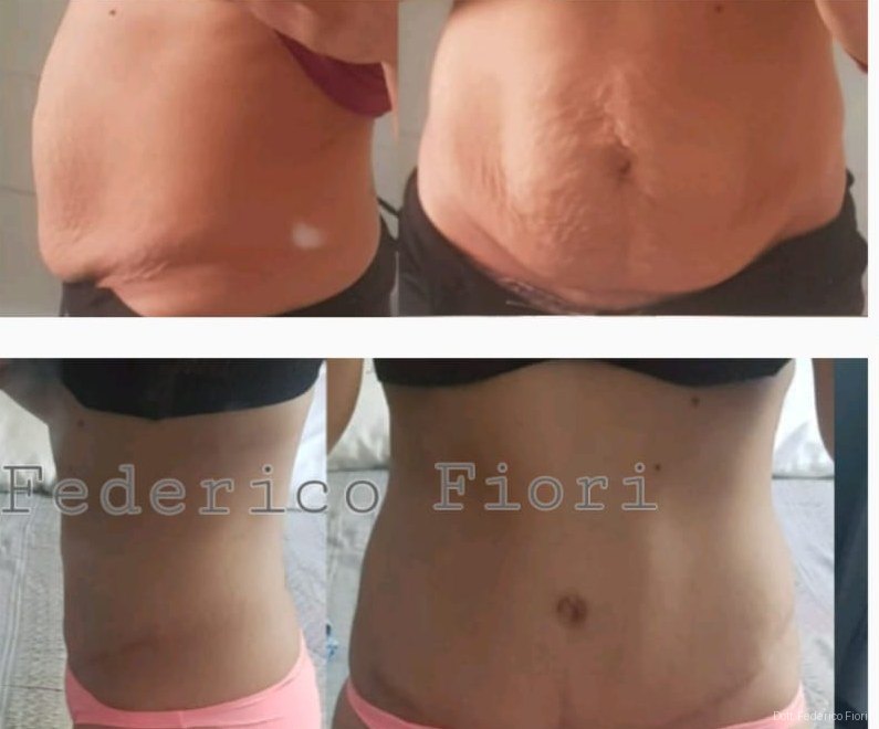
Before and after Rectus diastasis
Rectus diastasis
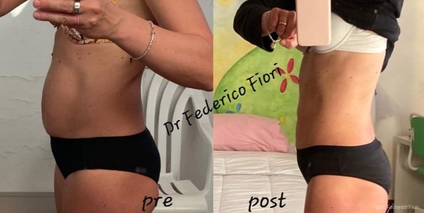
Before and after Rectus diastasis
Rectus diastasis
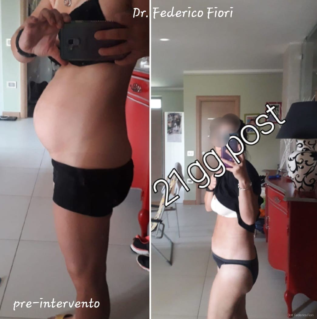
Before and after Rectus diastasis
Rectus diastasis
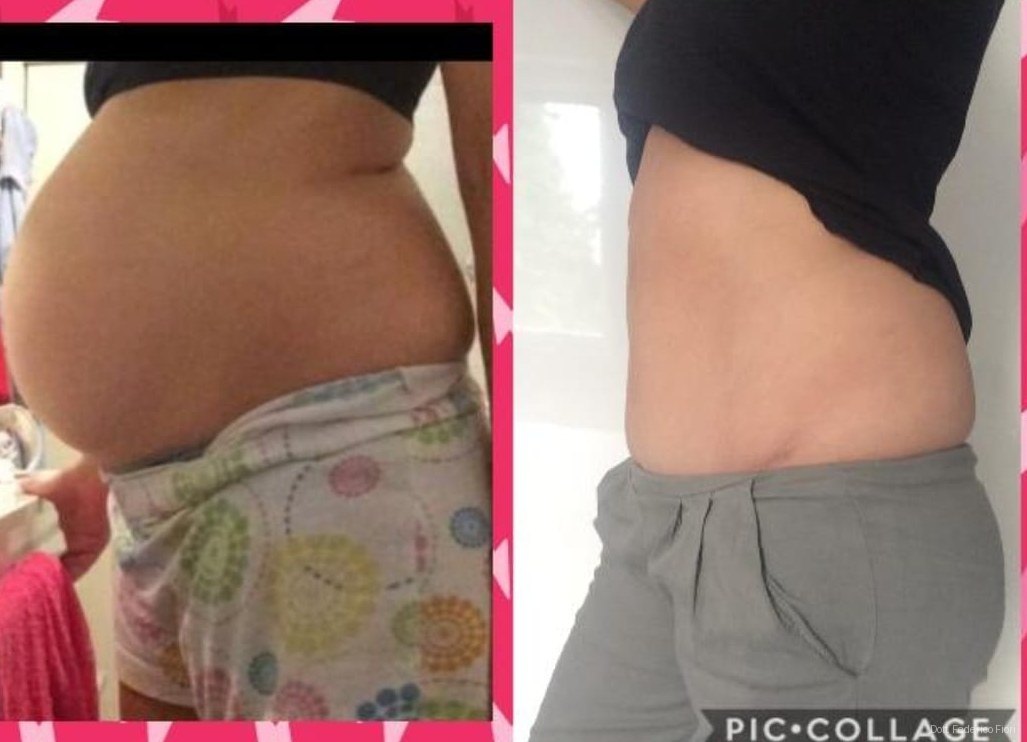
Before and after Rectus diastasis
Rectus diastasis
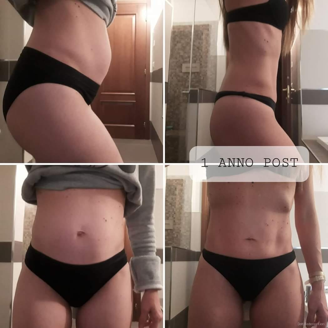
Before and after Rectus diastasis
Rectus diastasis
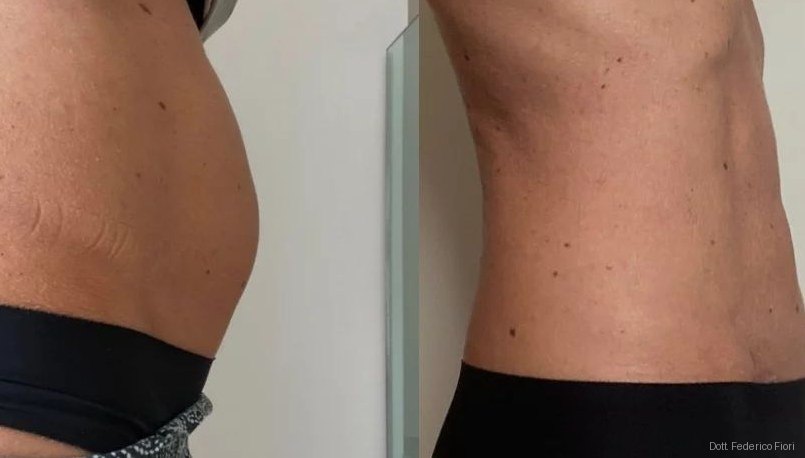
Before and after Rectus diastasis
Rectus diastasis
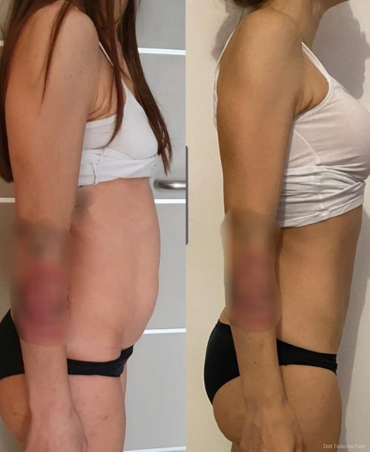
Before and after Rectus diastasis
Rectus diastasis
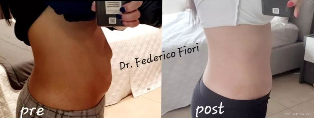
Before and after Rectus diastasis
Rectus diastasis
Before and after Rectus diastasis
Rectus diastasis
Before and after Rectus diastasis
Rectus diastasis
Before and after Rectus diastasis
Rectus diastasis
Before and after Rectus diastasis
Rectus diastasis
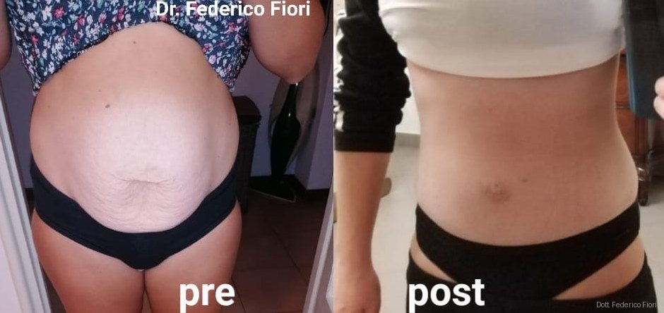
Before and after Rectus diastasis
Rectus diastasis
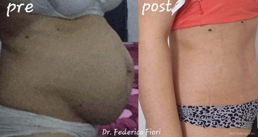
Before and after Rectus diastasis
Rectus diastasis
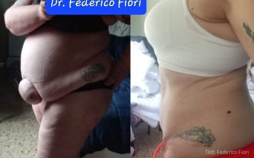
Before and after Rectus diastasis
Rectus diastasis
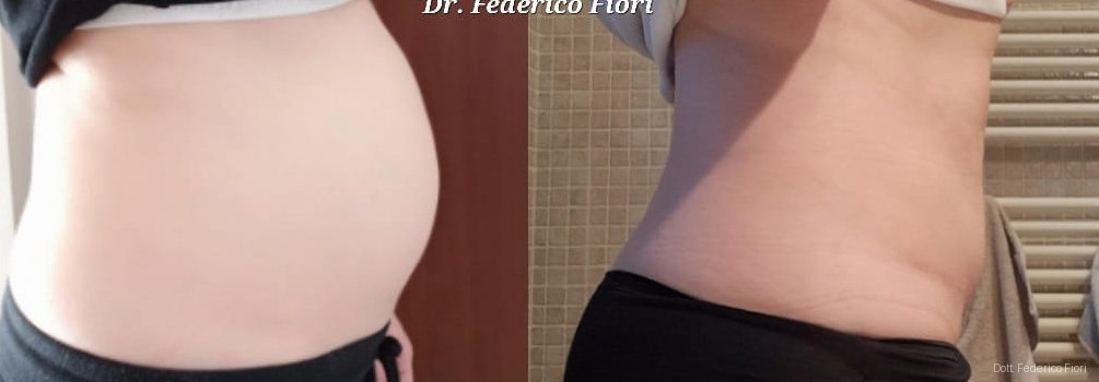
Before and after Rectus diastasis
Rectus diastasis
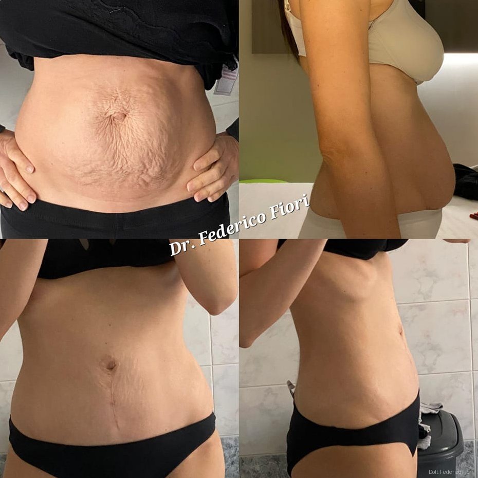
Before and after Rectus diastasis
Rectus diastasis
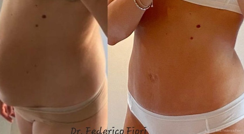
Before and after Rectus diastasis
Rectus diastasis
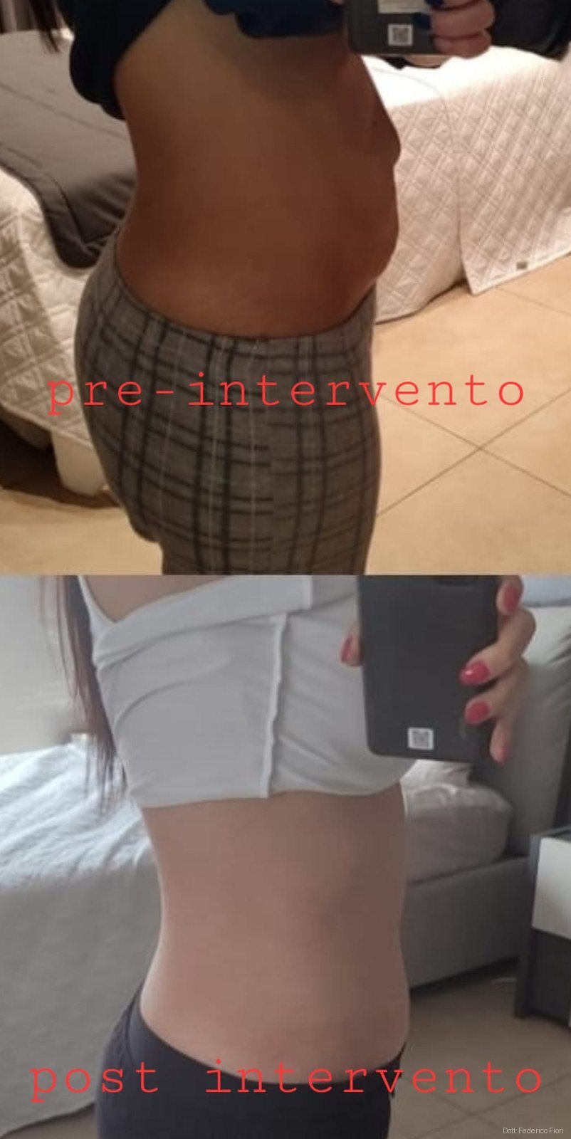
Before and after Rectus diastasis
Rectus diastasis
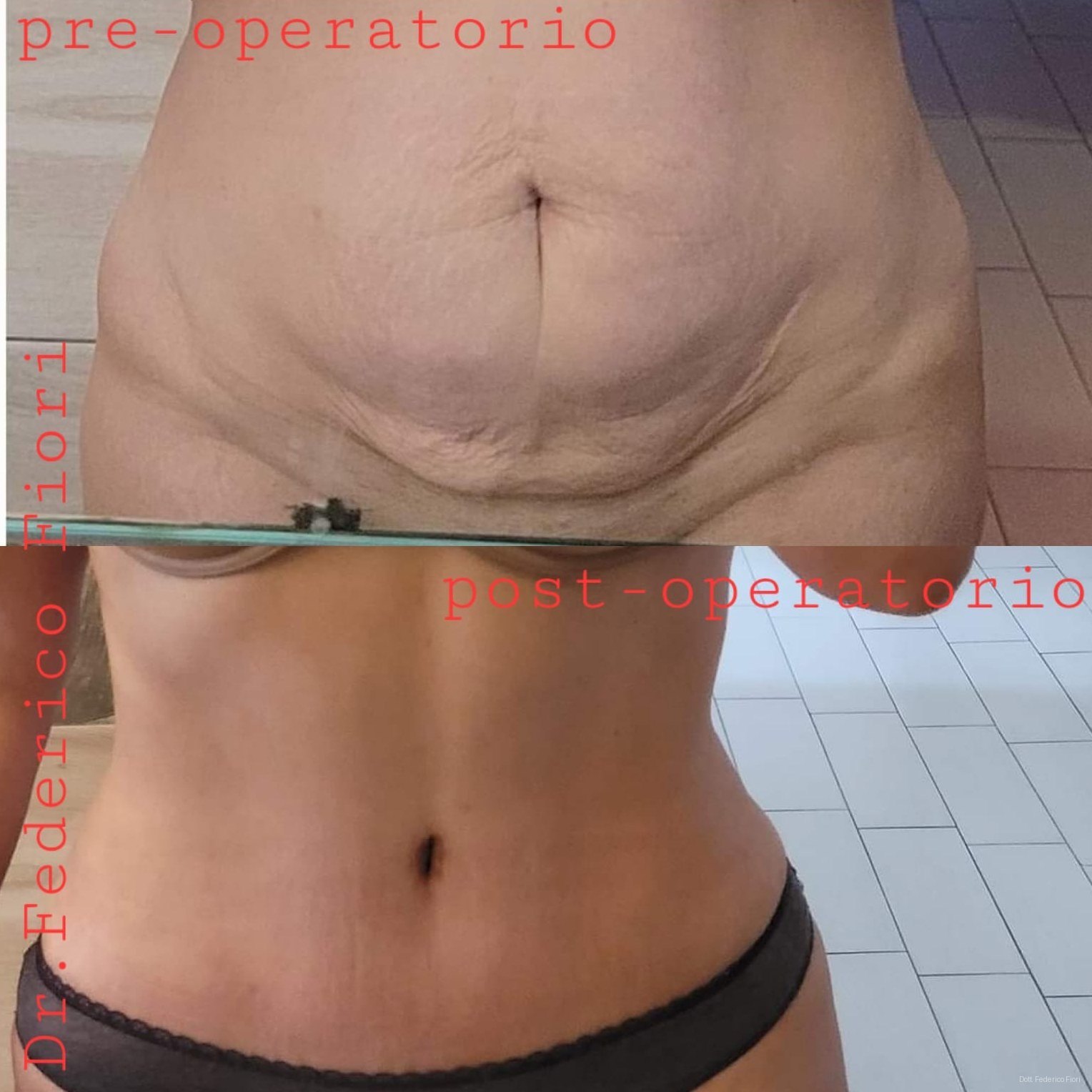
Before and after Rectus diastasis
Rectus diastasis
Before and after Rectus diastasis
Rectus diastasis
Before and after Rectus diastasis
Rectus diastasis
Before and after Rectus diastasis
Rectus diastasis
Before and after Rectus diastasis
Rectus diastasis
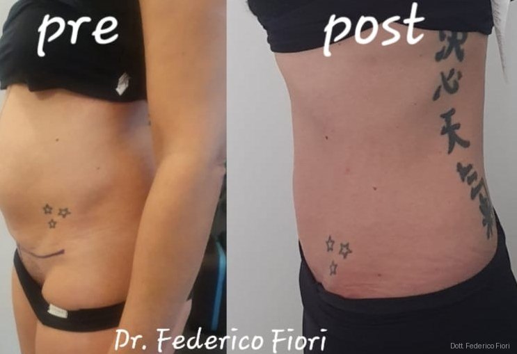
Before and after Rectus diastasis
Rectus diastasis
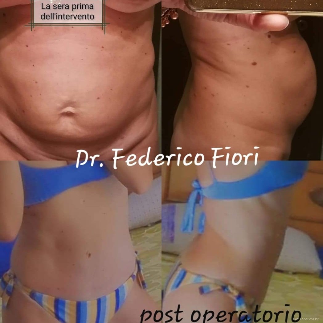
Before and after Rectus diastasis
Rectus diastasis
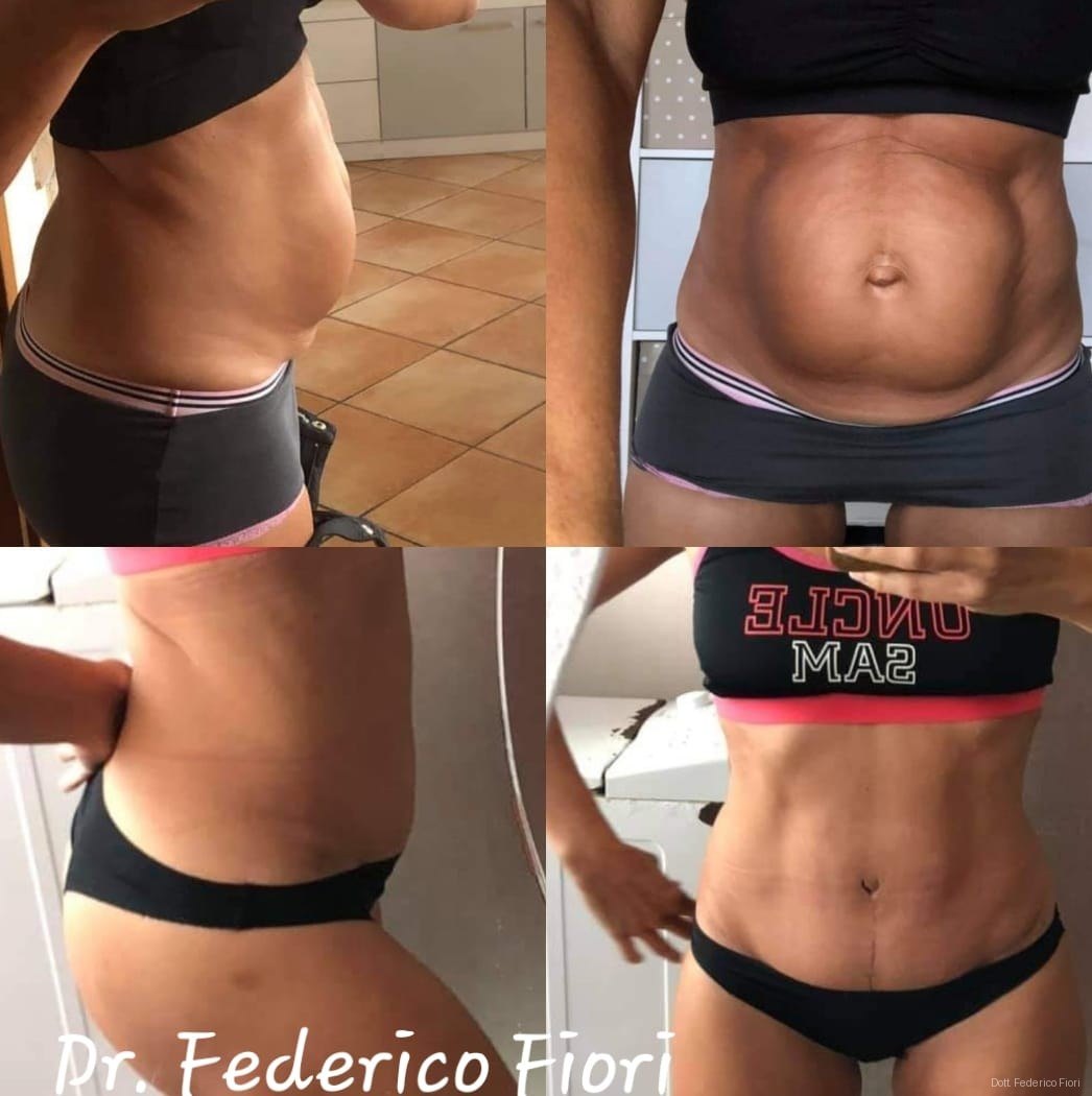
Before and after Rectus diastasis
Rectus diastasis
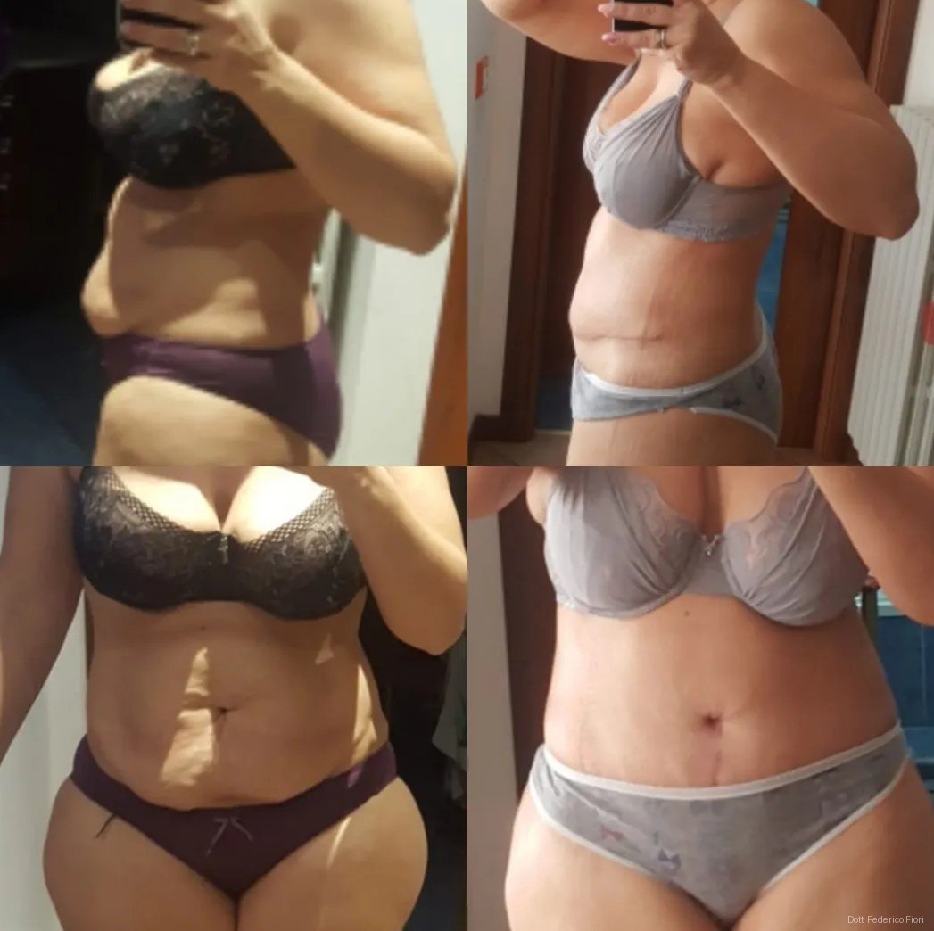
Before and after Rectus diastasis
Rectus diastasis
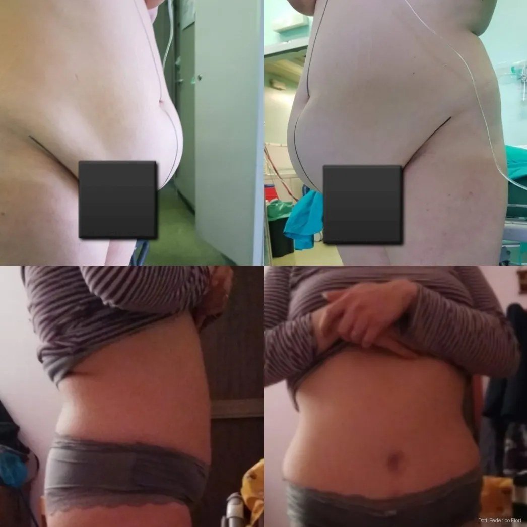
Before and after
Images published with specific written consent. Faces and identifying marks are not recognisable.
Frequently asked questions about results
- Do before and after images guarantee the same result in my case?
- No, and they should not be read that way. They document what was achieved in that specific case, with that technique and from that starting point. The result achievable in your case is established at the consultation, with clinical examination and imaging.
- How many months after surgery are the post-operative photographs taken?
- It is stated case by case. Generally the useful images are those from three to twelve months: before that, swelling is still present and scars are redder and more raised than they will eventually be.
- Did the people consent to publication?
- Yes. Every image is published under written consent specific to publication, separate from consent to surgery and revocable at any time.
- Why are faces not shown?
- Because they are not needed, and because a patient's photograph is health data. The images are cropped to the abdominal region and identifying features are masked.
- Will the scar stay as it looks in the images?
- In most cases it continues to improve. A scar matures over twelve to eighteen months, changing in color and in how raised it is. Site, tension, genetics, smoking and sun protection all play a part.
- Can I see other cases similar to mine?
- Yes, at the consultation. I show documentation of cases comparable to your situation, including images of partial outcomes: they help you understand the real limits of what is achievable.
Last updated: 2026-08-20 · Content by Dr. Federico Fiori