The TESAR Technique for Ventral Hernias, Incisional Hernias and Rectus Diastasis
TESAR — Totally Endoscopic Sublay Anterior Repair — repairs rectus diastasis and midline hernias, both primary and incisional, endoscopically, placing the mesh in the retromuscular plane. The access is anterior and the abdominal cavity is never opened: the wall is rebuilt by working between its own layers.
| Acronym | Totally Endoscopic Sublay Anterior Repair |
|---|---|
| Access | Anterior, endoscopic, extraperitoneal |
| Mesh position | Retromuscular (sublay) |
| Indications | Midline ventral and incisional hernias, rectus diastasis |
| First publication | J Laparoendosc Adv Surg Tech, 2019 |
What does TESAR stand for?
TESAR stands for Totally Endoscopic Sublay Anterior Repair: a fully endoscopic repair, with the mesh in the sublay position, through an anterior access. Each word of the acronym describes a specific technical choice: the operation is endoscopic, the mesh sits behind the muscle, and the access route is anterior.
After the first operations performed in 2018, I published it in 2019 in the Journal of Laparoendoscopic & Advanced Surgical Techniques. Since 2024 it has been included, alongside the other extraperitoneal endoscopic techniques, in the classification proposed in a systematic review published in Hernia.
How is the operation performed?
The operative sequence is as follows:
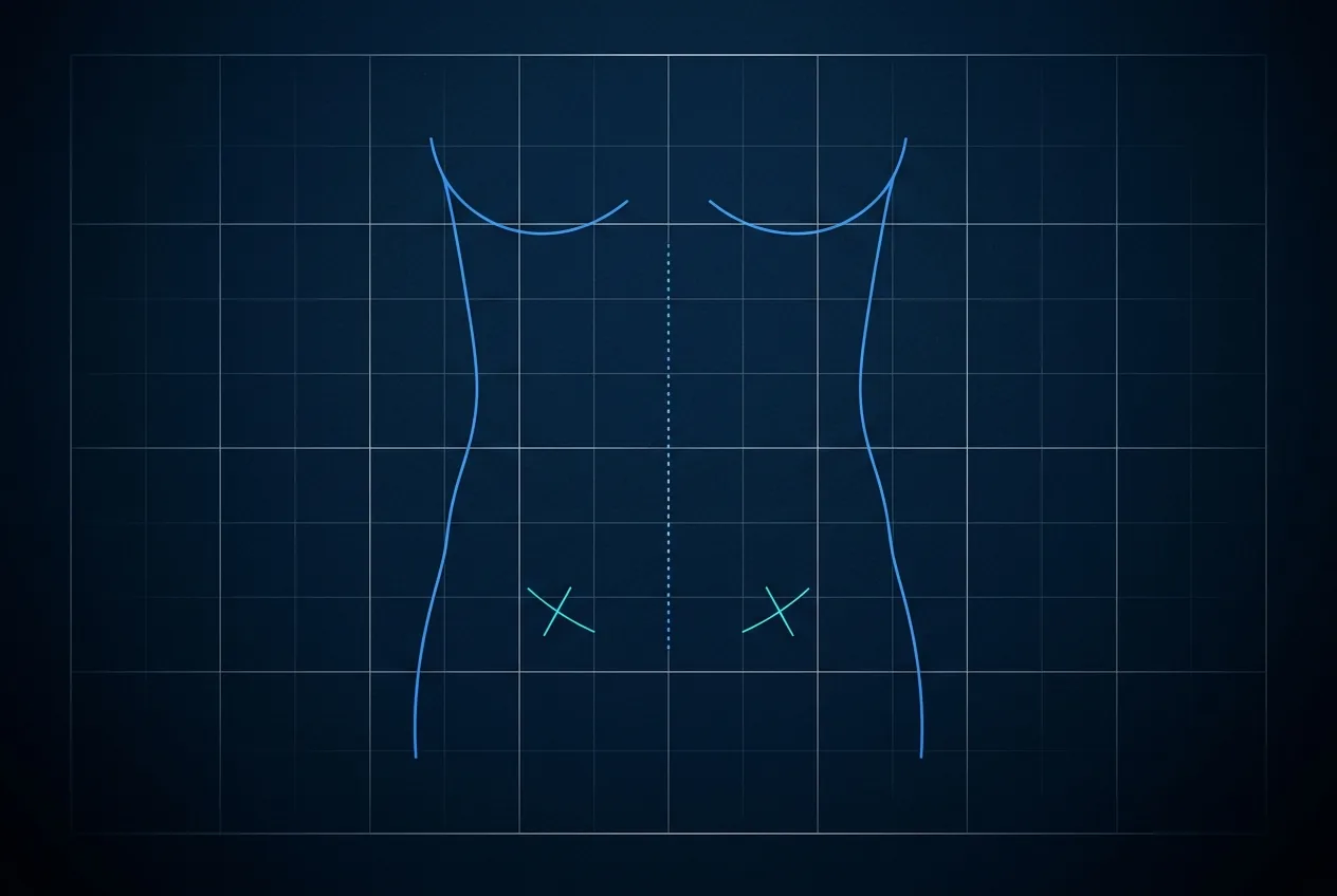
- a suprapubic access through small incisions;
- endoscopic pre-aponeurotic dissection — above the aponeurosis — to create the working space;
- incision of the medial edges of the anterior rectus sheaths;
- placement of the synthetic mesh in the retromuscular position, behind the rectus muscle;
- closure of the sheaths over the mesh, restoring the continuity of the midline.
All the work takes place outside the abdominal cavity: the operation is extraperitoneal. One technically relevant consequence follows — the mesh never comes into contact with the viscera.
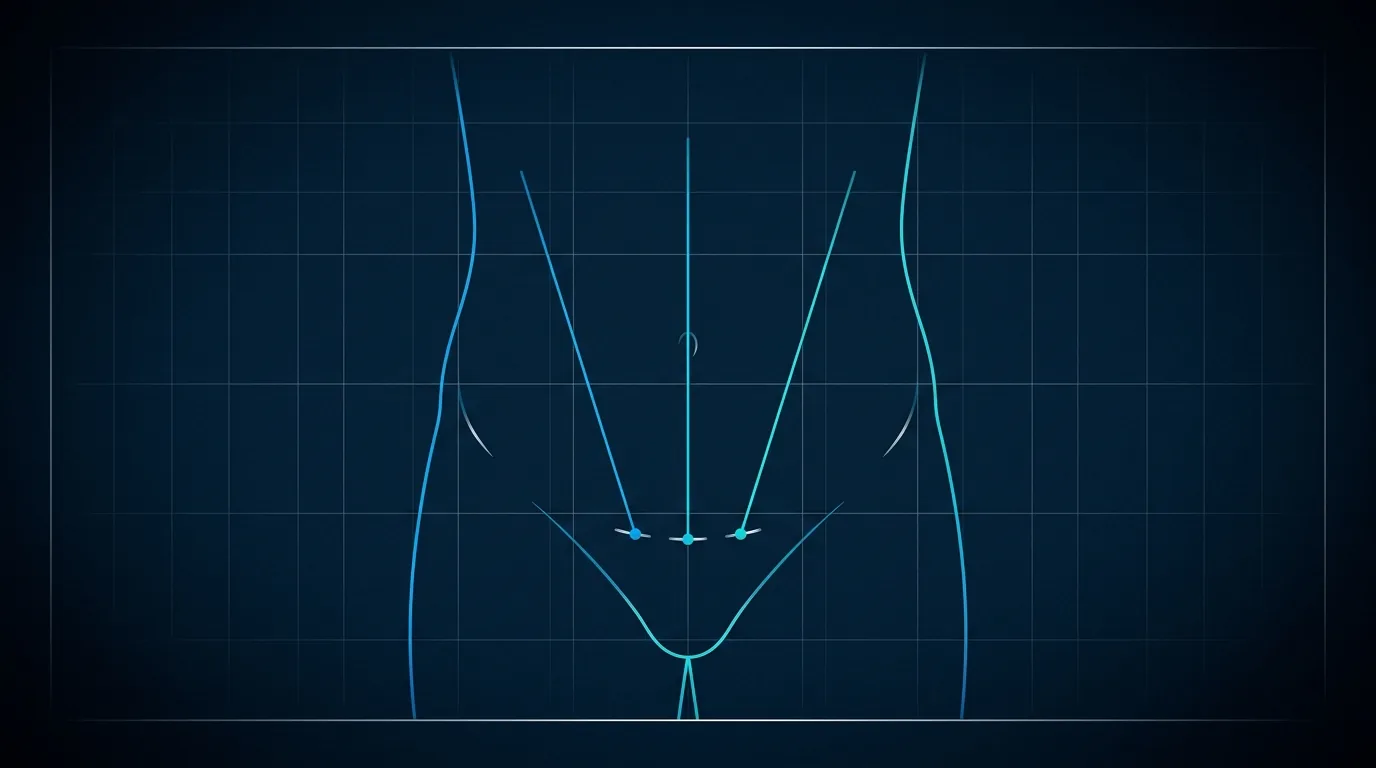
Other steps are added to these, and they do not serve to close the defect but to give the wall back its ability to work:
- the posterior plane can be plicated to increase the tension of the transverse muscles, which in diastasis have lost their load and are effectively re-tensioned;
- where indicated, the oblique muscles are addressed to reduce the circumference of the abdominal core;
- a transverse plication at the level of the umbilicus compensates, where necessary, for the vertical laxity of the fascia.
These are steps that concern function. Correcting shape, in this technique as in every minimally invasive technique, is not a primary aim: the improvement in shape is the consequence of properly restoring function.
How does it differ from other anterior approaches?
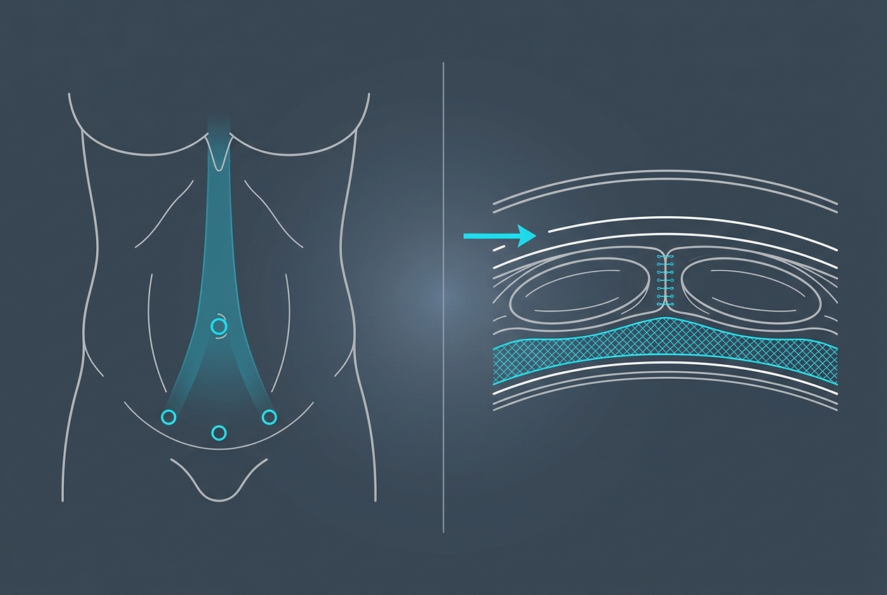
Historically, endoscopic techniques working through an anterior route place the mesh in the onlay position, that is, above the aponeurosis. Techniques that place the mesh retromuscularly, on the other hand, generally do so through a posterior route.
TESAR combines the two: an anterior route with a retromuscular mesh. That is the technical point that sets it apart, and the reason why, in the 2024 systematic review of extraperitoneal approaches, it is the only anterior technique classified with the mesh in the sublay position (Hernia 2024 — DOI 10.1007/s10029-024-03144-3).
What results has it produced over time?
The long-term outcomes of the first 120 consecutive operations were published in Hernia in 2026, with a median follow-up of 31 months and observations up to 81 months; 22 patients passed the five-year mark. Only patients whose follow-up we verified ourselves were included, through clinical examination or imaging: those who did not attend the check-up were excluded from the analysis, even though they had reported no problem. The series comprises 83 cases of diastasis with umbilical hernia, 39 incisional hernias and 15 other primary ventral hernias; 45.8% of the patients had already had abdominal surgery.
There were no intraoperative complications. Overall postoperative morbidity was 6.7%: six seromas (5%), none of which required a further operation, and two haematomas (1.6%), one of which required revision. There were no surgical site infections and no clinical recurrence was detected during the observation period. Median hospital stay was three days.
In functional terms the EuraHS-QoL score — the instrument that measures quality of life specific to abdominal wall surgery — went from a median of 57 before surgery to 0 at twelve months, across the 104 patients assessed. A lower score means a better condition: at one year patients reported the absence of the complaints they had before. The pain domain went from 17 to 0, the activity restriction domain from 26 to 0, the cosmetic domain from 14 to 0; the Carolinas Comfort Scale from 35.5 to 0. (Hernia 2026;30:305 — DOI 10.1007/s10029-026-03808-2 · PMID 42536212)
Managing the dead space
The most frequent complication of every technique involving subcutaneous undermining is seroma, the collection of fluid in the space created during the operation. Since 26 April 2023 the technique includes a specific modification to prevent it: a progressive-tension suture anchoring the subcutaneous tissue to the fascia along the midline, abolishing that space and distributing tension. The effect was measured by comparing patients operated on before and after its introduction: clinically relevant seromas went from six out of seventy-five to none out of forty-five.
Which conditions is it indicated for?
- midline ventral hernias, including umbilical and epigastric hernias;
- incisional hernias on a midline scar;
- rectus diastasis, isolated or associated with a hernia defect;
- situations in which reconstruction of the linea alba is part of the goal, not a side effect.
When is it not the right choice?
A technique is only as useful as its stated limits. TESAR is not the right answer when:
- the excess of skin and fatty tissue is such that it requires surgical remodelling of the soft tissues, which has to be planned together with the repair: in these cases no minimally invasive technique is indicated;
- the defect is very wide or involves a loss of domain that requires component separation techniques;
- particular local or general conditions make another approach preferable, whether minimally invasive or open, for instance in the presence of comorbidities;
- the clinical assessment concludes that no operation is needed: this too is an indication, and it must be stated as clearly as the others.
How does it fit into the morpho-functional approach?
TESAR is a technical tool. It is useful when the problem in front of us coincides with what this technique solves well: a midline defect in which the structure has to be rebuilt and the mesh has to go behind the muscle. When the picture is different — because the morphological component carries more weight and cannot be handled with a minimally invasive procedure, or because the defect is of another nature — the right choice has to be a different one.
It is the same logic described on the page about the morpho-functional approach: first the right question, then the technique.
Where has it been published and discussed?
Selected publications
- Totally Endoscopic Sublay Anterior Repair for Ventral and Incisional Hernias. Journal of Laparoendoscopic & Advanced Surgical Techniques, 2019. PMID 30807248 · DOI 10.1089/lap.2018.0807 First description of the TESAR technique.
- Surgical treatment of diastasis recti: the importance of an overall view of the problem. Hernia, 2021. DOI 10.1007/s10029-020-02252-0 Functional and morphological outcomes assessed with the EuraHS-QoL tool.
- Laparoendoscopic extraperitoneal surgical techniques for ventral hernias and diastasis recti repair: a systematic review. Hernia, 2024. PMID 39312025 · DOI 10.1007/s10029-024-03144-3 Classification of anterior and posterior extraperitoneal approaches.
- Totally Endoscopic Sublay Anterior Repair (TESAR) for diastasis recti, primary and incisional ventral hernias: long-term clinical outcomes and quality of life. Hernia 2026;30(1):305, 2026. PMID 42536212 · PMCID PMC13427856 · DOI 10.1007/s10029-026-03808-2 Long-term clinical outcomes and quality of life after TESAR, observational study.
Partial list, being updated. Every reference can be verified through PubMed or DOI.
Lectures and teaching
- Speaker at the congresses of the European Hernia Society (EHS)
- Speaker at the congresses of the Italian Association of Hospital Surgeons (ACOI)
- Live surgery and teaching for the SIC-ISHAWS National School of abdominal wall surgery
- Lectures at national and international courses and congresses on abdominal wall and minimally invasive surgery
- Public-education meetings for patients and health professionals
Glossary of minimally invasive abdominal wall acronyms
MILA: minimally invasive lipoabdominoplasty
Frequently asked questions about the TESAR technique
- What is the TESAR technique?
- TESAR (Totally Endoscopic Sublay Anterior Repair) is an extraperitoneal endoscopic technique for ventral and incisional hernias and for rectus diastasis: anterior access, mesh placed in the retromuscular position, no entry into the abdominal cavity.
- Who developed the TESAR technique?
- It was developed and described by Dr. Federico Fiori, who published the first description in 2019 in the Journal of Laparoendoscopic & Advanced Surgical Techniques.
- How does TESAR differ from other anterior endoscopic techniques?
- Other anterior techniques usually place the mesh above the aponeurosis (onlay). TESAR keeps the anterior access but positions the mesh behind the rectus muscle (sublay).
- Is TESAR also used for rectus diastasis?
- Yes: it is indicated for rectus diastasis, isolated or associated with a midline hernia, when the goal includes reconstruction of the linea alba.
- Does the mesh come into contact with the bowel?
- No. The operation is extraperitoneal: the mesh is placed behind the rectus muscle and does not come into contact with the viscera.
- Is TESAR always the best technique?
- No. It is one tool among others. Very large defects, tissue loss, or a significant excess of skin and fat call for different solutions, to be planned during the assessment.
- How long is the hospital stay?
- In the published series of the first 120 operations the median stay was three days, with a range from two to ten. The time needed to return to daily and sporting activities is defined case by case, according to the defect treated and the reconstructive steps performed.
- What is the risk of recurrence?
- In the published follow-up — a median of 31 months, with 22 patients beyond five years — no clinical recurrence was detected across the first 120 consecutive operations, which are those with follow-up verified by us.
Last updated: 2026-08-20 · Content by Dr. Federico Fiori